What Is Sonography And How The Images Are Obtained?
Sonography is one of the pivotal Imaging Tests that is scheduled that helps physicians to diagnose and treat medical conditions through viewing the ultrasound images of organs, tissues or blood flow of inside the body. 3D/4D Sonography In Delhi is available in a limited number of Imaging Centre that is well equipped with modern technologies and expert technicians who understand the use of it. Arun Imaging is the centre located in Vivek Vihar, which offers excellent services and features to people.
Sonography Defined –
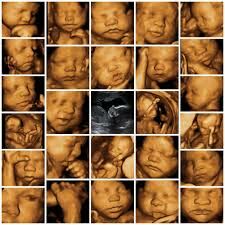
The sizes and shapes of structures, tissues and organs can be seen in a sonogram image. It can be useful to detect medical tissues such as gallbladder disease or gallstones, kidney stones or kidney disease, liver disease, appendicitis, ovarian cysts, ectopic pregnancies, uterine growths or fibroid, and other conditions. The most common use of a sonogram is to monitor the development of the uterus and fetus during pregnancy.
The Attainment Of Images –
An ultrasound machine, a transducer and coupling gel are the basic requirements of performing this test. The coupling gel is applied to the area which is to be diagnosed as it creates a bridge between the transducer and the skin. The transducer generates ultrasound which is directed through the skin in the body. The organs reflect back the ultrasound waves back and then the echoes are sent to the ultrasound machine. The electrical signals get converted into an image and displayed on the monitor.
We have the most reliable Imaging Centre In Delhi where we offer amiable services without overcharging our customers. You can get your appointment fixed with us over a call.

Comments
Post a Comment